
CAT scan (computer axial tomography):
A CAT scanner emits a series of narrow beams through the human body as it
moves through an arc. Inside the CAT scanner there is an X-ray detector which can see hundreds
of different levels of density. This data is transmitted to a computer, which builds up a 3-D
cross-sectional picture of the part of the body and displays it on the
screen.
 Structural MRI (Magnetic Resonance Image):
Diffusion-Tensor MRI (DTI):
Diffusion Tensor Magnetic Resonance Imaging, or DT-MRI, is a technology
that measures the random motion of hydrogen atoms within water molecules
in all three dimensions, non-invasively, and in vivo. DT-MRI adds to conventional MRI the capability of measuring the random motion of water molecules, referred to as diffusion.
 EEG (electroencephalograph):
The recording machine changes the electrical signals into patterns that can be seen on a computer. It looks like wavy lines and this data can help diagnose seizures or other illnesses.

PET scan (Positron emission tomography):
A PET scan is an imaging test that uses a
radioactive substance called a tracer to look for disease in the body. A PET scan uses a small amount of radioactive material (tracer). The
tracer is given through a vein (IV), most often on the inside of your
elbow. The tracer travels through your blood and collects in organs and
tissues. This helps the radiologist see certain areas of concern more
clearly. How long the test takes depends on what part of the body is being scanned.

fMRI (functional MRI):
fMRI is based on the same technology as magnetic resonance imaging (MRI) -- a noninvasive test that uses a strong magnetic field and radio waves to create detailed images of the body. But instead of creating images of organs and tissues like MRI, fMRI looks at bloodflow in the brain to detect areas of activity. These changes in blood flow, which are captured on a computer, help doctors understand more about how the brain works.

phMRI (pharamacological MRI):
the use of fMRI to map spatiotemporal patterns of brain activity induced by pharmacological agents, has provided a robust and flexible tool to resolve brain circuits and mechanism-specific functional changes produced by selective intervention in different neurotransmitter systems in vivo.
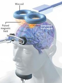 TMS (transcranial magnetic stimulation):
TMS uses a magnet instead of an electrical current to activate the brain. An electromagnetic coil is held against the forehead and short electromagnetic pulses are administered through the coil. The magnetic pulse easily passes through the skull, and causes small electrical currents that stimulate nerve cells in the targeted brain region. And because this type of pulse generally does not reach further than two inches into the brain, scientists can select which parts of the brain will be affected and which will not be. The magnetic field is about the same strength as that of a magnetic resonance imaging (MRI) scan.
Sources:
Source 2
Source 3
No comments:
Post a Comment